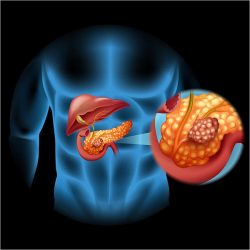
PANCREATIC CANCER
The pancreas is a large gland behind the stomach and next to the duodenum and small intestine. The pancreas does two main things: it releases powerful digestive enzymes into the small intestine to help digest food, and it releases the hormones insulin and glucagon into the bloodstream. These hormones help the body control how to use food for energy.
Pancreatic cancer is one of the most aggressive cancers and its incidence has been increasing in recent decades. It is more common in men than women and is mainly seen in patients over 60 years of age, with an increase in younger patients in recent years. It is the fourth leading cause of cancer death.
Its exact etiology is not completely known. Smoking and the consumption of various foods such as animal fat, animal protein and red meat are implicated. There are also a number of hereditary diseases that are associated with pancreatic cancer and constitute 10% of cases. Such diseases that predispose to the development of pancreatic cancer are hereditary pancreatitis, cystic fibrosis, familial breast cancer and familial pancreatic cancer.
The two oncogenes that have been found to be associated with pancreatic cancer are ras and c-erbb-2, which were found to be activated in 75 and 20% of cases respectively.
In contrast, the oncogenes fos and myc are expressed in a small percentage of patients (10%) and weakly, and this fact itself makes their use in clinical practice unlikely.
It was found that prognosis is not affected by the expression of oncogenes, but mainly by the stage of the disease.
Pancreatic adenocarcinoma is the most common form of pancreatic cancer and accounts for 85% of cases. Rarer forms are cystadenocarcinoma and tumors from the endocrine part of the pancreas. In 2/3 of cases it is located in the head of the pancreas and the rest concerns the body and tail of the organ.
Pancreatic cancer metastasizes hematogenously mainly to the liver and lungs and more rarely to other organs (bones, adrenal glands), lymphogenously to regional lymph nodes and then to tissues in the peritoneum and neighboring organs.
In most cases, the diagnosis of pancreatic cancer is delayed and this is due to the fact that the symptoms are often atypical and develop slowly. Cancer of the head of the pancreas usually manifests with weight loss, obstructive jaundice, deep epigastric pain and vomiting. Also rarely, the appearance of diabetes mellitus may be the first manifestation of cancer.
In pancreatic cancer, an increased tumor marker CA 19-9 is usually detected in the blood serum. For its diagnosis, there are many imaging methods: ultrasound, CT (computed tomography) and spiral CT, CT angiography, MRI (magnetic resonance imaging), MRCP, ERCP (endoscopic retrograde cholangiopancreatography), PET (positron emission tomography), endoscopic ultrasound, CT-guided or endoscopic ultrasound-guided biopsy (FNA). The above tests help, in addition to the diagnosis, to decide whether the tumor is resectable (vascular infiltration, presence of metastases in the liver and lungs).
In cases where the cancer is resectable, the treatment of choice is surgery (pancreatoduodenectomy-Whipple), which has very good results. Complementary chemotherapy is often required. Before starting the surgery, it is advisable to precede with a diagnostic laparoscopy.
This extensive surgery may be necessary in a few cases, but it is also dangerous and drastically changes the lifestyle. After a total pancreatectomy, a person will develop diabetes. He must change his diet, his daily routine and will have to take insulin for the rest of his life. Removing the pancreas will also reduce the body’s ability to break down food and absorb its nutrients. Taking pancreatic enzymes by mouth is essential in these cases. Without insulin injections and digestive enzymes, a person without a pancreas cannot survive. However, with the right medication, a person can live without a pancreas.
A special clinical condition is locally advanced pancreatic cancer.
The tumor has spread outside the pancreas, into the duodenum, bile duct, and other tissues surrounding the pancreas, surrounding and sometimes strangulating large blood vessels and nerves, and has also spread to lymph nodes, but not to other parts of the body.
Here too, the same diagnostic tools are used to see if it is resectable.
Preoperative endoscopic stent placement, according to some studies, is either not beneficial, or increases the overall rate of postoperative morbidity and mortality, or increases the risk of wound infection.
These patients should immediately undergo induction treatment with:
• Chemotherapy or
• Concurrent chemotherapy and radiotherapy
with the aim of both treating the symptoms caused by the cancer in order to extend the survival of the patients and the rare but real possibility of reducing the tumor to such an extent that it can be surgically removed.
The treatment regimen followed depends on the patient’s clinical condition and his ability to tolerate an aggressive drug regimen which can often present significant side effects.
It is important to remember that the more aggressive the therapeutic approach, the greater the likelihood of achieving our goal of controlling the disease.
The oncologist-pathologist’s obligation is to judge which of the patients are able to undergo aggressive treatment without negatively affecting their quality of life and which are not able to receive such treatment.
Once the treatment regimen is completed, imaging is performed with:
• Computed tomography
• Magnetic angiography of the splenoportal axis
• PET/CT.
• If there is HYPOSTAGING and no detection of systemic METASTASES in the liver, lungs or peritoneum, then the patient is considered a candidate for surgical curative resection.
Percutaneous placement of 125I with the help of a CT scan or their direct placement with surgery helps in the necrosis of cancer cells. Placement of an intraarterial catheter usually in the gastroduodenal artery for direct infusion of chemotherapeutic agents can shrink or delay the spread of the disease.
Sublimation of tumors with radio frequencies, radio waves, laser, IRE and helium Knife can also prolong and provide quality of life.


